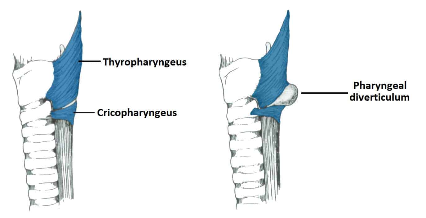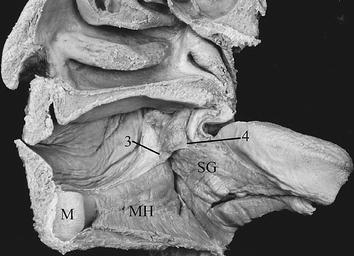


Es el músculo más alto de los tres constrictores faríngeos. Longitudinal muscles constitute the inner muscular layer and play a. El músculo constrictor faríngeo superior es un músculo de la faringe. It arises from the sides of the cricoid cartilage and the thyroid cartilage. Superior constrictor muscle Middle constrictor muscle Inferior constrictor muscle. The function of this muscle is to facilitate food propagation towards the esophagus by constricting the wall of pharynx during deglutition. It is the thickest of the three outer pharyngeal muscles. The inferior pharyngeal constrictor is a muscle of the pharynx that, together with the superior and middle pharyngeal constrictors, forms the posterior and lateral walls of laryngopharynx. Conclusion: This study clarified that the superior pharyngeal constrictor muscle came into contact with the buccinator muscle without intervention of the bone and originated from the mandible and root of the tongue. The inferior pharyngeal constrictor muscle is a skeletal muscle of the neck. Muscles originating from these four parts merged immediately after the origin and were aligned transversely towards the posterior pharyngeal wall. The superior constrictor muscle is located anterior to the prevertebral muscles and posterior to the buccinator muscle, from which it is separated by the pterygomandibular raphe. The muscle at the glossopharyngeal part originated from the root of the tongue in all cases. The deep aspect of the superior constrictor muscle is separated from the prevertebral fascia by a thin layer of retropharyngeal fat. Morphology of the origin of the muscle at the mylopharyngeal part could be divided into two types: type I, tip of the origin of the muscle on the mylohyoid line and type II, tip of the origin of the muscle away from the mylohyoid line. Morphology of the origin of the muscle at the buccopharyngeal part could be divided into three types: type I, membranous morphology from superior to inferior areas type II, membranous only in superior area and type III, complete lack of membrane. Results: The superior pharyngeal constrictor muscle at the pterygopharyngeal part originated between the pterygoid hamulus of the sphenoid bone and the posterior margin of the medial pterygoid plate to form the most superior area of the pharyngeal wall. The superior pharyngeal constrictor muscle was observed from the facial and oral sides. Methods: 37 cadavers of Japanese adults that had been fixed in 10% formaldehyde and stored at the Department of Anatomy in Tokyo Dental College were used. The pharyngeal branch of the vagus nerve via the pharyngeal plexus supplies the superior pharyngeal constrictor muscle.Objectives: To clarify the details morphology of the superior pharyngeal constrictor muscle in humans during ingestion movement, we examined the macroscopic characteristics of the pterygopharyngeal, buccopharyngeal, mylopharyngeal and glossopharyngeal parts of the muscle. The constrictors contract upon the bolus and transmit it down inside the esophagus.Immediately, the bolus of food is attached in the pharynx: The stylopharyngeus muscle separated its lower margin from the middle constrictor.Below the base of the skull, a crescent gap occurs above the muscle in which the auditory tube and the levator as well as tensor veli palatini muscles arise.

The highest fibres are attached to the pharyngeal tubercle of the occipital bone and the lowest fibres are covered from the middle constrictor. The pharyngeal constrictors include the following muscles: Superior pharyngeal constrictor - the most superior constrictor muscle, located in the oropharynx. Insertion: Pharyngeal raphe, pharyngeal tubercle Nerve: Vagus nerve Action: Swallowing Description: The Superior constrictor (Constrictor pharyngis superior) is a quadrilateral muscle, thinner and paler than the other two.The muscular fibres spread out backwards, and are attached into a raphe outspreading down the posterior wall of the pharynx within the median plane, for the most part.Glossopharyngeus part – From the mucous membrane of the Hoor of the mouth.Mylopharyngeus part – From the mylohyoid line of the mandible.Buccopharyngeus part – From the pterygomandibular raphe. The Superior constrictor (Constrictor pharyngis superior) is a quadrilateral muscle, thinner and paler than the other two.Pterygopharyngeus part – From the inferior half of the posterior border of the medial lamina of the pterygoid process.superior pharyngeal constrictor muscle arises continuously:


 0 kommentar(er)
